AI use in Medical Diagnosis and Prognosis of Cancer
Allison Calle, Jin Chao Chen, Edison Mak, Cielo Raymundo
Abstract
Background:
The diagnosis of cancer is a slow and complex process that restricts access to many people due to its cost. Cancer is an unpredictable disease that normally takes a great amount of time and resources to diagnose. People who are financially struggling or have a hard time getting around also have a hard time obtaining the proper diagnoses and treatments in time.
Objective:
Our objective is to develop and test an implementation of artificial intelligence that can be used to accurately and efficiently diagnose cancer.
Design:
Our proposed solution for the implementation of artificial intelligence to diagnose patients involves the development of a body scanner device. This device, with the help of advanced AI technology, would scan a patient and provide results and feedback within minutes.
Results:
The use of artificial intelligence in the diagnosis of cancer would greatly benefit patients and medical practitioners alike. The machine would be more efficient, accurate, and cost-effective while being unaffected by factors that would typically reduce the accuracy of diagnoses, such as misinformation and human error. The usage of AI-powered machines and processes may improve the physical and financial accessibility of such important medical procedures.
Literature Review
Over the years, the amount of diseases have increased and become too complex to diagnose and prevent. As a result, doctors are spending more time and resources examining patients trying to ensure accurate diagnoses. As the medical field grows, doctors are expected to implement and search for advanced and efficient methods to support their work and ease their load.
The evolution of artificial intelligence is unceasing. Its marvelous performance has been seen in many fields, including gaming and art. With studies presenting its great potential, the medical field can bloom with its integration. AI can provide support to the medical field, ranging from medical imaging to drug design. Its stability, accuracy, machine learning and utilization of big data promises a potential automated omnitool if given enough resources and time. Most recently, advanced AI – powered machines have provided the ability for doctors to do a full body scan on their patients within minutes. No longer need lengthy procedures or even risky surgeries for doctors to know what is wrong. This enables doctors to save valuable time and focus on patients after care more.
Cancer detection using AI has been one of the most breakthrough areas. Cancer is one of the most unpredictable diseases in existence. It can be deadly when not detected as soon as possible. In the past, doctors have found it incredibly difficult to diagnose or predict cancer and how it would spread. “AI may be used to find patterns in enormous volumes of medical data, aiding in disease prediction and prevention before symptoms appear”(Umapathy et al., 2023). Having doctors use these AI supported machines helps them have a second pair of eyes and helps them identify abnormalities or potential issues that might go undetected at first sight. It enables patients’ confidence in doctors as they want to be seen thoroughly when going to the hospital.
Using pattern recognition is probably one of the major factors that helped improve AI predictions. Having AI remember all these imaging and past movements can help predict future movement of current patients more accurately without second guessing. For instance, “a novel AI-based approach was developed to accurately predict the preoperative stage of rectal cancer via computed tomography (CT) images.”(Yan et al., 2023, p. 4) With this it also made the development of a disease prognosis more efficient, having all these past diagnoses helps machines detect diseases earlier which is beneficial for survival and cure rates. “AI-based radiomics has played an increasingly important role in the clinical
decision-making of HCC patients, providing new technical guarantees for prediction, diagnosis, and prognostication.”(Chen et al., 2024) With these predictive algorithms on diseases it can help those patients foresee potential risks before they even appear, which helps save lives.
As the usage of AI in the medical field continues to evolve so do its opposing factors. Apart from it being too expensive for most healthcare providers to implement and maintain these AI systems, there are concerns of data, lack of trust and lack of empathy in these systems. AI needs large sets of ranging data to be able to recognize patterns and accurately provide diagnosis and prognosis, the data must be completely unbiased. The diagnostic accuracy of clinicians decreases significantly when they are provided with predictions from a systemically biased AI model. A systemically biased AI model is a model trained on data that are consistently flawed, such as data that misdiagnoses certain patient subpopulations. (Jabbour et. al, p1) To get this unbiased data the existing concerns of using sensitive patient data security and privacy to train these AI models must disappear. The concerns for this data surround the questions of if patients have consented for their health information to be used and if there is a chance of data breaching and are the patients aware of data vulnerability.
These concerns surrounding data sensitivity and ethical attainability also go into the lack of trust and empathy that medical professionals have in how AI comes to its conclusions. This specific trust concern stems from “the black box” phenomenon. The black box nature refers to medical professionals not knowing how an AI model came to the conclusion it did (Rainmanee, p1). The term “black box” comes from the nested linear structures that these models have (Deshpande et. al, p844). Doctors and medical professionals cannot trust this technology if it is not transparent. The lack of trust and empathy from the patients stems from their understanding that by integrating AI into the health process, this means that the doctors will be cut out from the middle and all the communication will be made solely through AI models.
The benefits of using AI in the medical field such as accuracy and early detection to diagnose cancer and provide an accurate prognosis, as well as its ability to make solid decisions in stressful environments cannot be overlooked. The controversies surrounding the usage of AI in medical environments are mostly regarding the way it’s being trained and used, as well as the inaccuracies of the models themselves. In other words, AI as a fundamental concept has immense potential which can be achieved by reducing its flaws.
The flaws in AI’s implementation in the medical field only serve as a reason to accelerate development of more precise AI models and improved practices such as transparency about where data is sourced and how it is implemented. Moreover, AI is soon to hatch from its chaotic blackbox nature. Normally, the user–in this case, a doctor–wouldn’t understand how the AI makes its conclusions and can’t double check them, resulting in distrust. This creates a need for an AI with opaque innerworkings, an AI that can explain its claims, an explainable artificial intelligence (XAI). It already exists! A comparison experiment between XAI with and without SHAP (a technique that explains the output of any machine learning model) shows that XAI with SHAP performed about 20% better. (Zhang, 4.2)
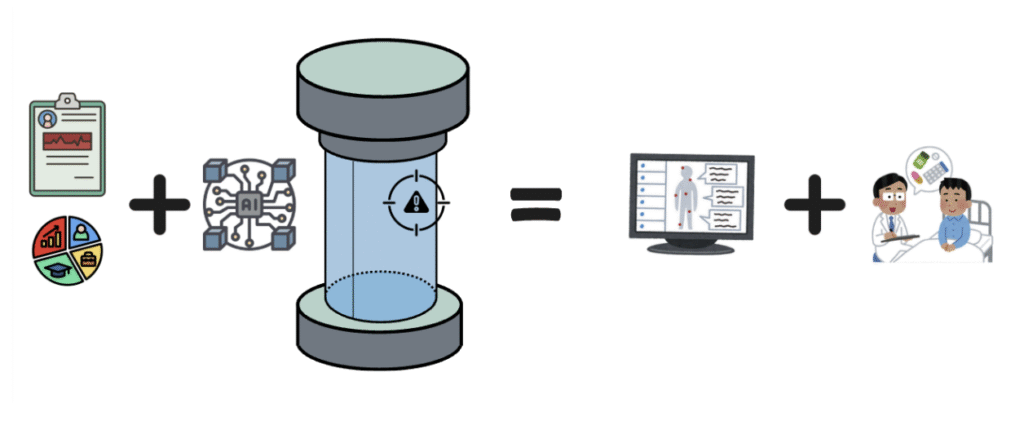
“Onco Imaging” could be the solution: It is the comprehensive collection of the previously stated leading technology, its energy efficient, has large memory, gives rapid analysis and is highly explainable. Structurally, “OI” is a large upright cylindrical machine in which a person is able to step inside. This machine is then connected to a control panel, which includes the operator console, vitals monitors, and computer equipment that controls the scanner of the machine. It uses advanced technology to scan the human body and along with an AI that uses archived data of cancer patients, it is able to diagnose and give a prognosis of cancer. This AI works by using algorithms that learn from large amounts of archived cancer patient data sets to recognize patterns and make predictions which typically require human intelligence and time-consuming procedures. Doctors then can read, analyze and deliver these processed conclusions.
Methods
To test the accuracy and efficiency of Onco Imaging, a crossover design trial was executed. A total of fifty participants were recruited via outreach at health fairs and community centers. The participants were all randomly chosen based on a demographics questionnaire and an interview focusing on their concerns of the possibility of having cancer. Their ages ranged from 18 to 63, they all had different backgrounds/ethnicities and economic statuses. A total of 30 medical professionals were recruited through medical conferences that focused on oncology, this group included nurses, doctors and social workers. After the participants were chosen, half of them were randomly assigned to go through the control group first (p=25) and the other half through the experimental group first (p=25). The caregivers were also randomly split between the control (m=20) and experimental (m=10) groups.
The control group consists of the participant going through the current regular process of being diagnosed. This includes even being dismissed by care providers or going through the full process of physical exams, biopsies, laboratory and imaging testing. The experimental group consists of the participant going through the usage of OI and doctors analyzing findings. The experimental group consists of taking the participants to the OI. They are first brought to the information station, where they provide their background information and family medical history. Once that is complete they step into the OI where they are scanned entirely for a few mins. OI captures their biometric data that helps show the medical staff whether or not they have cancer.
Once the participants for the control and experimental group were finalized, they each went through both processes. Both the participants and caregivers were kept unaware of the results from the other group in order to not influence their expectations and findings. At the end of both processes the results were compared.
All the medical data used to train OI was voluntarily provided directly by either the patient or by their immediate family members
Anticipated Results
The goal we are trying to achieve is to provide a way for improving a doctor’s efficiency and accuracy when dealing with cancer. Currently, the main problem we have is slow response time, limited decision making and organization, which lead to inaccurate results. This procedure can result in a patient not receiving their results up to several weeks due to manual data processing and lab experiment.
Onco Imaging will only take about 7 minutes to scan and give results there and then, fully automatic. Turning the wait time from days or weeks to minutes. Each device makes its conclusion by applying algorithms with data based off of countless past cases and in-time scanner readings. Due to this decision making will not depend on imperfect induction of doctors but on reliable and precise knowledge.
To see if OI was reliable we conducted a study to show the diagnosis between caregivers and OI for similarities. In the study it was shown that there were a number of participants that were not diagnosed correctly.
| Group | # of Caregivers | # of Participants in the experiment | # of participants that were not diagnosed with cancer | # of participants that were diagnosed cancer | # of Participants correctly diagnosed by OI | # of Participants correctly diagnosed by doctors |
| Control | 20 | 25 | 3 | 22 | 19 | 17 |
| Experimental | 10 | 25 | 5 | 20 | 17 | 9 |
The results shown above demonstrate that participants diagnosed by OI had a higher accuracy of diagnosis compared to those diagnosed by doctors.This is where the gap of falsely diagnosed patients with cancer is covered by OI. This machine takes in past information from past cases that help it precisely diagnose a patient. Cancer can be a tricky disease and can be hard to diagnose in the early stages. Some of the early stages of cancer aren’t visibly shown which makes it harder for medical staff to diagnose early.
Broader Impact
With cancer being the leading cause of death worldwide, “accounting for nearly 10 million deaths in 2020, or nearly one in six deaths” (World Health Organization, 2025). This project will enable doctors and medical professionals to detect cancer in its early stages. Early detection before symptoms appear is crucial due to it drastically increasing the rate of survival of the patient as it provides the most treatment options (Bourgeois, 2024) and is when treatments are likely to be the most effective. Given the existing disparity and bias in the medical diagnosis of cancer across the world, “Racialized minorities bear a disproportionate cancer burden, including some of the highest rates of cancer-related morbidity and lowest rates of survival” (Bourgeois et. al, 2024). This technology will empower those who have been overlooked for many years, due to multiple factors like ethnicity, race, economic status, and more. This innovative project will not only help decrease the total global deaths every year by providing earlier diagnosis and improving a person’s prognosis. But, it will also revolutionize the entire medical industry by leading a new age of diagnosing diseases that have huge disparities leading to greater health equity.
References
Bo, Z., Song, J., He, Q., Chen, B., Chen, Z., Xie, X., Shu, D., Chen, K., Wang, Y., & Chen, G. (2024). Application of artificial intelligence radiomics in the diagnosis, treatment, and prognosis of hepatocellular carcinoma. Computers in Biology and Medicine, 173, 108337. https://doi.org/10.1016/j.compbiomed.2024.108337
Deshpande et. al (2022) Explainable Artificial Intelligence–A New Step towards the Trust in Medical Diagnosis with AI Frameworks: A Review Department of Electronics & Telecommunication, Lavale, Symbiosis Institute of Technology, Symbiosis International https://www.proquest.com/docview/3200120308
Eskandar, K., et al. “Artificial Intelligence in Healthcare: Explore the Applications of AI in Various Medical Domains, Such as Medical Imaging, Diagnosis, Drug Discovery, and Patient Care.” Series of Medical Science
Jabbour, S., Fouhey, D., Shepard, S., Valley, T. S., Kazerooni, E. A., Banovic, N., Wiens, J., & Sjoding, M. W. (2023). Measuring the Impact of AI in the Diagnosis of Hospitalized Patients: A Randomized Clinical Vignette Survey Study. JAMA : The Journal of the American Medical Association, 330(23), 2275–2284. https://jamanetwork.com/journals/jama/fullarticle/2812908
Rainmanee, W. (2025). AI Ethics: Should you trust AI with your medical diagnosis?. Journal of Integrative and Innovative Humanities Volume 5, Issue 1 https://so07.tci-thaijo.org/index.php/DJIIH/article/view/6207/4822
Zhang, YiMing, et al. “Applications of Explainable Artificial Intelligence in Diagnosis and Surgery.” MDPI, 2022
Bourgeois, A., Horrill, T., Mollison, A. et al. (2024) Barriers to Cancer Treatment for People Experiencing Socioeconomic Disadvantage in High-income Countries: A Scoping Review. BMC Health Serv Res 24, 670. https://doi.org/10.1186/s12913-024-11129-2
Smedley BD, Stith AY, Nelson A. (2003) Unequal Treatment: Confronting Racial and Ethnic Disparities in Health Care. Institute of Medicine (US) Committee on Understanding and Eliminating Racial and Ethnic Disparities in Health Care. National Academies Press (US)
World Health Organization. (2025) Cancer WHO https://www.who.int/news-room/fact-sheets/detail/cancer